Difference between revisions of "Template:Article of the week"
Shawndouglas (talk | contribs) m (Image size) |
Shawndouglas (talk | contribs) (Updated article of the week text) |
||
| (326 intermediate revisions by the same user not shown) | |||
| Line 1: | Line 1: | ||
<div style="float: left; margin: 0.5em 0.9em 0.4em 0em;">[[File: | <div style="float: left; margin: 0.5em 0.9em 0.4em 0em;">[[File:Fig4 Auer CytometryPartA2018 93-7.jpg|240px]]</div> | ||
'''"[[Journal:ChromaWizard: An open-source image analysis software for multicolor fluorescence in situ hybridization analysis|ChromaWizard: An open-source image analysis software for multicolor fluorescence in situ hybridization analysis]]"''' | |||
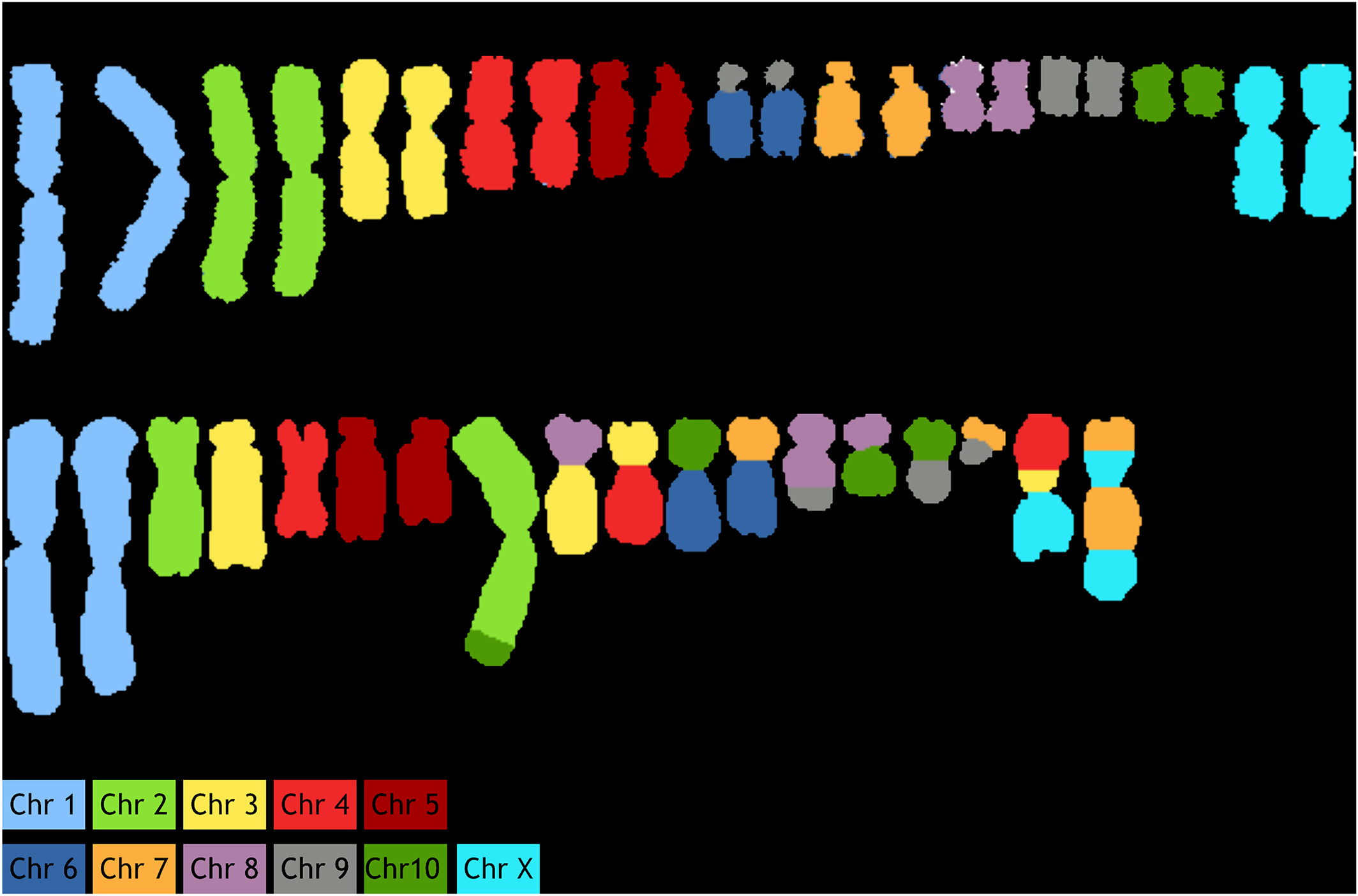
Multicolor image analysis finds its applications in a broad range of biological studies. Specifically, multiplex [[wikipedia:Fluorescence in situ hybridization|fluorescence ''in situ'' hybridization]] (M‐FISH) for chromosome painting facilitates the analysis of individual chromosomes in complex metaphase spreads and is widely used to detect both numerical and structural aberrations. While this is well established for human and mouse [[wikipedia:Karyotype|karyotypes]], for which species sophisticated software and analysis tools are available, other organisms and species are less well served. Commercially available software is proprietary and not easily adaptable to other karyotypes. Therefore, a publicly available open-source software that combines flexibility and customizable functionalities is needed. Here we present such a tool, called “ChromaWizard,” which is based on popular scientific image analysis libraries (OpenCV, scikit‐image, and NumPy). We demonstrate its functionality on the example of primary Chinese hamster (''Cricetulus griseus'') fibroblasts metaphase spreads and on Chinese hamster ovary cell lines, known for their large number of chromosomal rearrangements. ('''[[Journal:ChromaWizard: An open-source image analysis software for multicolor fluorescence in situ hybridization analysis|Full article...]]''')<br /> | |||
<br /> | <br /> | ||
''Recently featured'': [[ | ''Recently featured'': | ||
: ▪ [[Journal:Haves and have nots must find a better way: The case for open scientific hardware|Haves and have nots must find a better way: The case for open scientific hardware]] | |||
: ▪ [[Journal:CytoConverter: A web-based tool to convert karyotypes to genomic coordinates|CytoConverter: A web-based tool to convert karyotypes to genomic coordinates]] | |||
: ▪ [[Journal:Implementing a novel quality improvement-based approach to data quality monitoring and enhancement in a multipurpose clinical registry|Implementing a novel quality improvement-based approach to data quality monitoring and enhancement in a multipurpose clinical registry]] | |||
Revision as of 22:58, 24 February 2020
Multicolor image analysis finds its applications in a broad range of biological studies. Specifically, multiplex fluorescence in situ hybridization (M‐FISH) for chromosome painting facilitates the analysis of individual chromosomes in complex metaphase spreads and is widely used to detect both numerical and structural aberrations. While this is well established for human and mouse karyotypes, for which species sophisticated software and analysis tools are available, other organisms and species are less well served. Commercially available software is proprietary and not easily adaptable to other karyotypes. Therefore, a publicly available open-source software that combines flexibility and customizable functionalities is needed. Here we present such a tool, called “ChromaWizard,” which is based on popular scientific image analysis libraries (OpenCV, scikit‐image, and NumPy). We demonstrate its functionality on the example of primary Chinese hamster (Cricetulus griseus) fibroblasts metaphase spreads and on Chinese hamster ovary cell lines, known for their large number of chromosomal rearrangements. (Full article...)
Recently featured:


