File:Fig1 Deroulers DiagnosticPath2013 8.jpg
Original file (1,210 × 530 pixels, file size: 404 KB, MIME type: image/jpeg)
Summary
| Description |
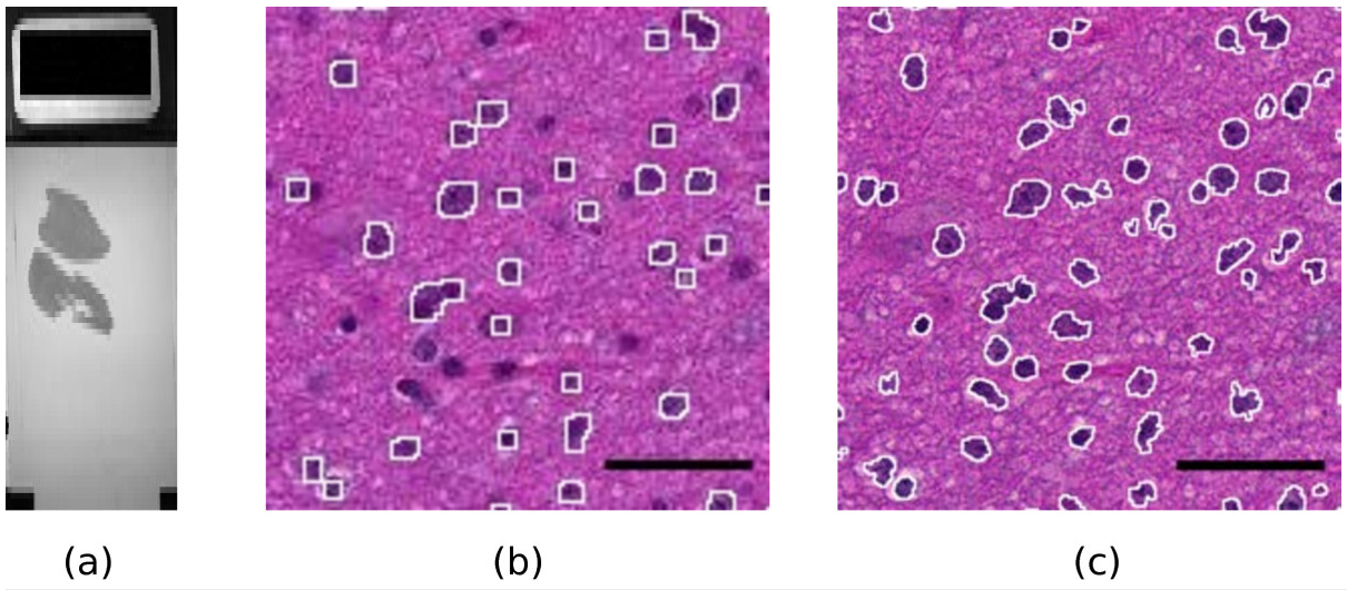
Figure 1. A sample slide. (a): macroscopic view of the whole slide (the black rectangle on the left is 1x2 cm). (b,c): Influence of the magnification on the quality of results. (b): a portion of the slide scanned at magnification level 10x. The white contours show the result of an automatic detection of the dark cell nuclei with the ImageJ software. A significant fraction of the cell nuclei is missed and the contours are rather pixelated. (c): the same portion of the slide scanned at magnification 40x. The white contours show the result of the same automatic detection. Almost all cell nuclei are detected and the shapes of the contours are much more precise. Scale bar: 4 μm. |
|---|---|
| Source |
Deroulers, Christophe; Ameisen, David; Badoual, Mathilde; Gerin, Chloé; Granier, Alexandre; Lartaud, Marc (2013). "Analyzing huge pathology images with open source software". Diagnostic Pathology 8: 92. doi:10.1186/1746-1596-8-92. ISSN 1746-1596. http://www.diagnosticpathology.org/content/8/1/92. |
| Date |
2013 |
| Author |
Deroulers, Christophe; Ameisen, David; Badoual, Mathilde; Gerin, Chloé; Granier, Alexandre; Lartaud, Marc |
| Permission (Reusing this file) |
|
| Other versions |
Licensing
|
|
This work is licensed under the Creative Commons Attribution 2.0 License. |
File history
Click on a date/time to view the file as it appeared at that time.
| Date/Time | Thumbnail | Dimensions | User | Comment | |
|---|---|---|---|---|---|
| current | 21:53, 26 September 2015 | | 1,210 × 530 (404 KB) | Shawndouglas (talk | contribs) |
You cannot overwrite this file.
File usage
The following page uses this file:


