File:Fig4 Fraggetta Diagnostics21 11-10.png

Original file (2,588 × 2,676 pixels, file size: 2.32 MB, MIME type: image/png)
Summary
| Description |
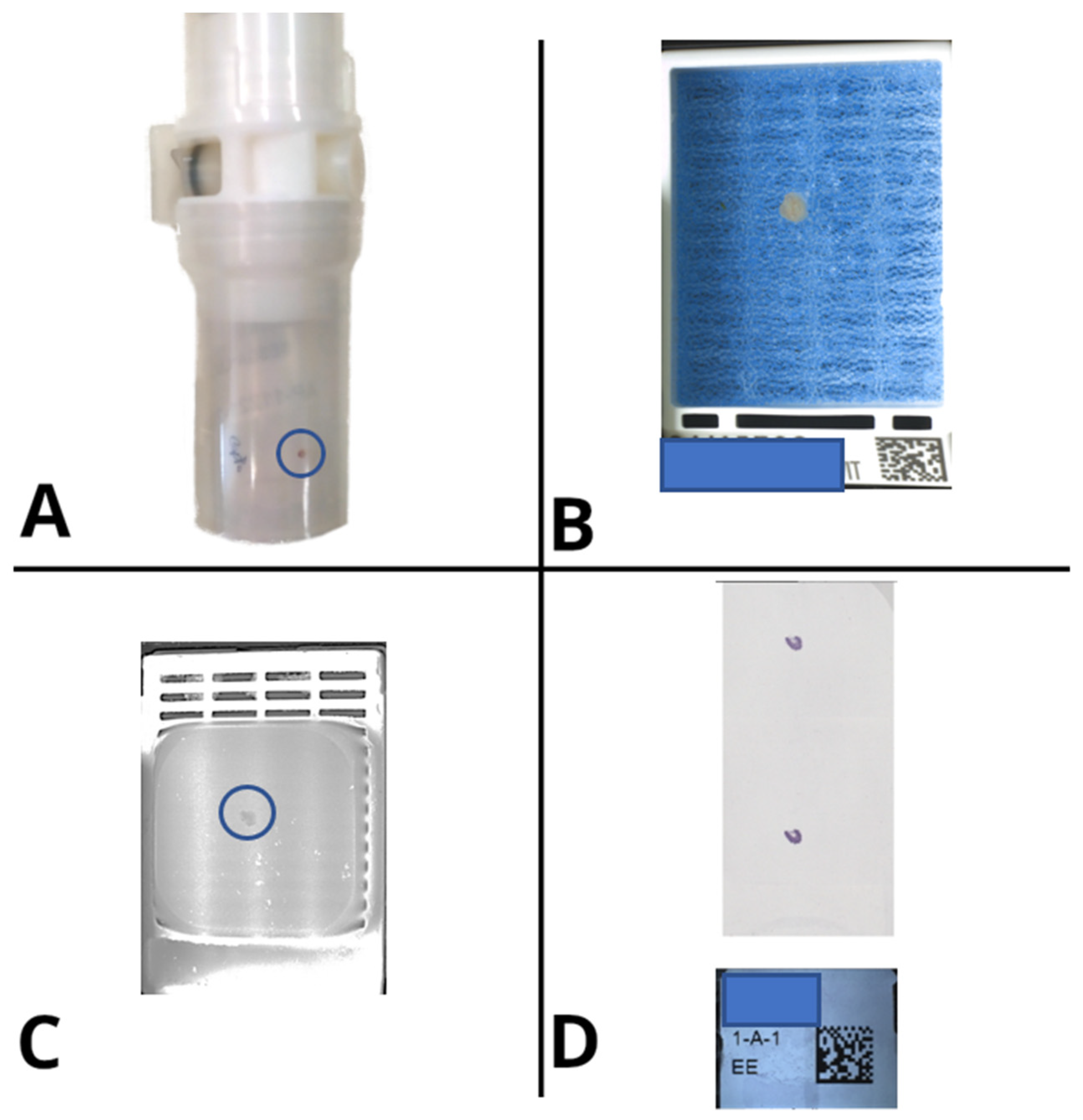
Figure 4. Digital pictures taken at each step of the life of the specimen and respective cassettes fully document the flow of tissue in the lab, allowing global traceability and high-resolution error tracking. A. Specimen container as it is received; B. Cassette at grossing, before closing its lid; C. Surface of the FFPE block after microtome sectioning; D. Macro picture of the glass slide after staining. |
|---|---|
| Source |
Fraggetta, F.; Caputo, A.; Guglielmino, R.; Pellegrino, M.G.; Runza, G.; L'Imperio, V. (2021). "A survival guide for the rapid transition to a fully digital workflow: The Caltagirone example". Diagnostics 11 (10): 1916. doi:10.3390/diagnostics11101916. |
| Date |
2021 |
| Author |
Fraggetta, F.; Caputo, A.; Guglielmino, R.; Pellegrino, M.G.; Runza, G.; L'Imperio, V. |
| Permission (Reusing this file) |
|
| Other versions |
Licensing
|
|
This work is licensed under the Creative Commons Attribution 4.0 License. |
File history
Click on a date/time to view the file as it appeared at that time.
| Date/Time | Thumbnail | Dimensions | User | Comment | |
|---|---|---|---|---|---|
| current | 18:28, 1 February 2022 |  | 2,588 × 2,676 (2.32 MB) | Shawndouglas (talk | contribs) |
You cannot overwrite this file.
File usage
The following page uses this file:


