File:Fig2 Larobina Tomography23 9-5.png
Original file (2,236 × 1,104 pixels, file size: 546 KB, MIME type: image/png)
Summary
| Description |
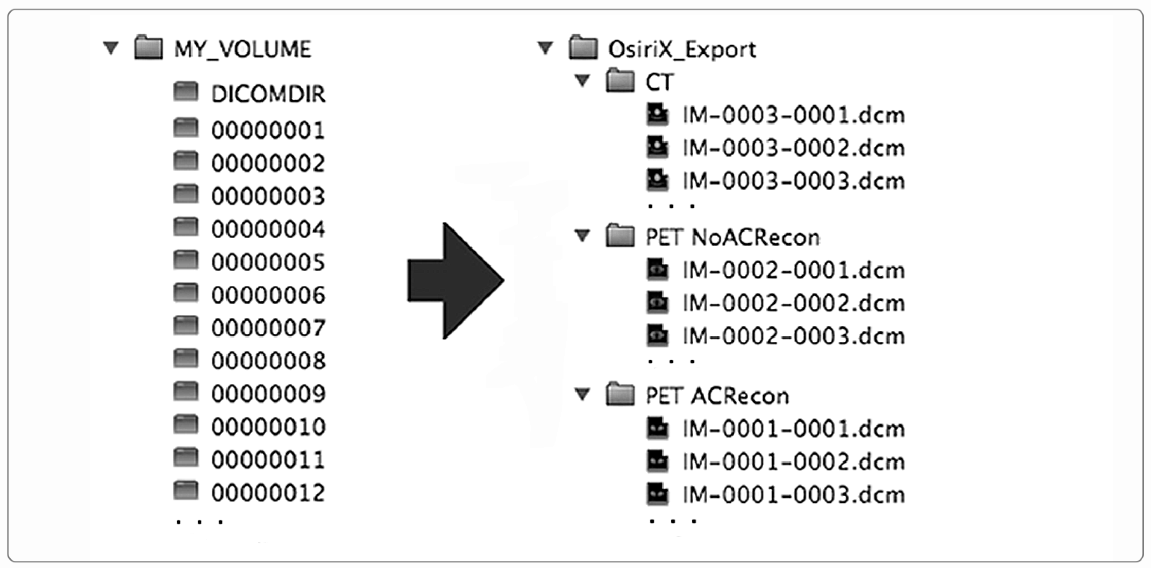
Figure 2. On the left, the DICOMDIR flat image files organization in the case of a patient who underwent a whole-body PET/CT diagnostic study. On the right, the corresponding image file organization using the hierarchical folder tree option (patient-study-series) in the OsiriX Viewer export utility. The PET study was reconstructed with and without attenuation correction (AC). According to the standard, DICOMDIR image filenames are no more than eight characters without any extension. |
|---|---|
| Source |
Larobina, M. (2023). "Thirty years of the DICOM standard". Tomography 9 (5): 1829-1838. doi:10.3390/tomography9050145. |
| Date |
2023 |
| Author |
Larobina, M. |
| Permission (Reusing this file) |
|
| Other versions |
Licensing
|
|
This work is licensed under the Creative Commons Attribution 4.0 License. |
File history
Click on a date/time to view the file as it appeared at that time.
| Date/Time | Thumbnail | Dimensions | User | Comment | |
|---|---|---|---|---|---|
| current | 18:29, 9 October 2023 | | 2,236 × 1,104 (546 KB) | Shawndouglas (talk | contribs) |
You cannot overwrite this file.
File usage
The following page uses this file:


