File:Fig1.1 Davis FrontBioinfo2022 40.jpg
Original file (699 × 704 pixels, file size: 208 KB, MIME type: image/jpeg)
Summary
| Description |
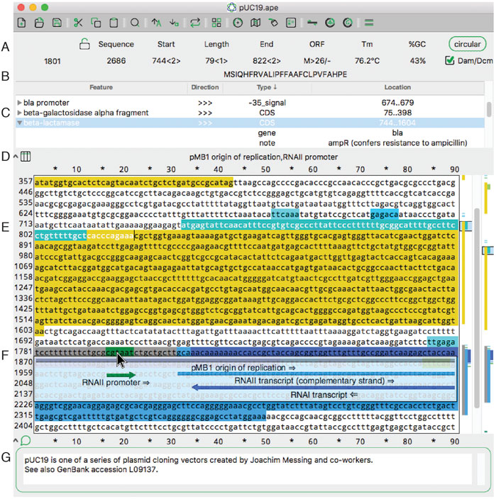
Figure 1. The main sequence editing window of ApE. (A) The top section of the window shows basic properties of the sequence and selected region. (B) The top section also shows the translation of the selected region. (C) The next pane shows a table of sequence features. Clicking on the arrowhead expands the description of the feature. (D) The next pane shows a list of all features under the mouse pointer (here, hovering over (F)). (E) The central region of the window contains the text of the sequence, with features highlighted in color. To the right is a vertical representation of these features in the currently displayed region and the scrollbar. On the far right is a representation of all of the features in the sequence. (F) When activated, the X-ray window shows a floating window containing a graphical representation of the line of text under the mouse pointer. (G) The bottom of the window shows an editable sequence comment. |
|---|---|
| Source |
Davis, M. W.; Jorgensen, E.M. (2022). "ApE, A Plasmid Editor: A freely available DNA manipulation and visualization program". Frontiers in Bioinformatics 40: e55. doi:10.1093/nar/gkr1288. |
| Date |
2022 |
| Author |
Davis, M. W.; Jorgensen, E.M. |
| Permission (Reusing this file) |
|
| Other versions |
Licensing
|
|
This work is licensed under the Creative Commons Attribution 4.0 License. |
File history
Click on a date/time to view the file as it appeared at that time.
| Date/Time | Thumbnail | Dimensions | User | Comment | |
|---|---|---|---|---|---|
| current | 18:29, 16 June 2023 | | 699 × 704 (208 KB) | Shawndouglas (talk | contribs) |
You cannot overwrite this file.
File usage
The following 2 pages use this file:


