File:Fig1 Larobina Tomography23 9-5.png
Original file (3,145 × 992 pixels, file size: 153 KB, MIME type: image/png)
Summary
| Description |
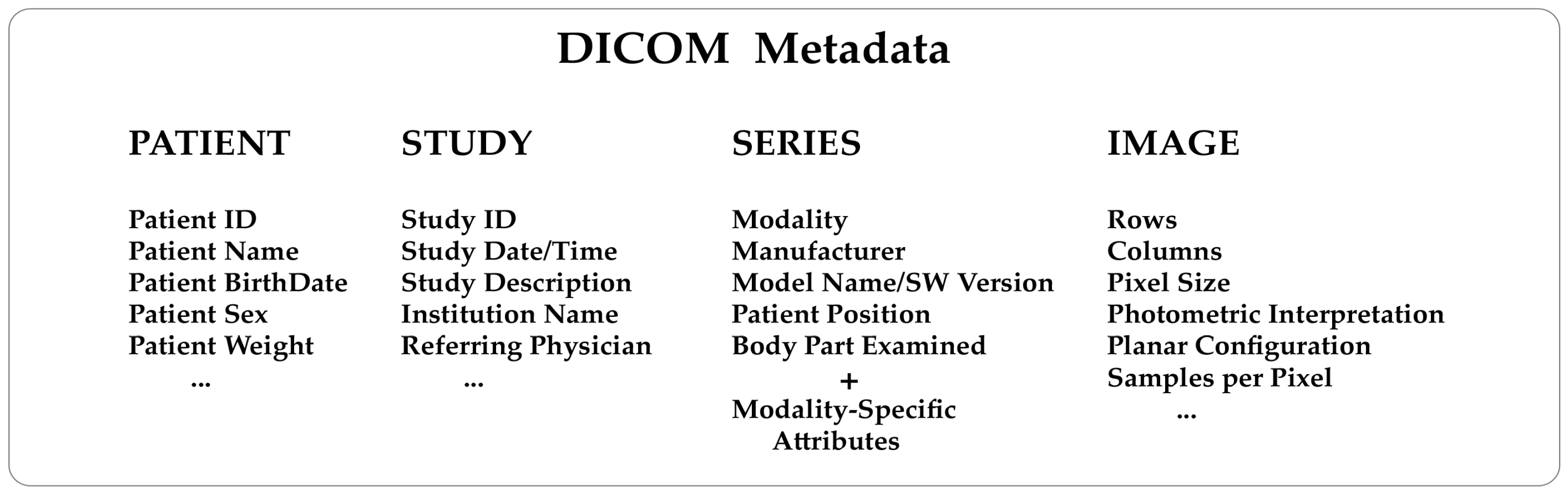
Figure 1. DICOM image metadata contains detailed information to identify and describe the main entities of the imaging workflow: Patient, Study, Series, and Images. The Patient data section essentially includes a unique patient identifier, the patient’s name, date of birth, sex, and data such as the patient’s weight, which is required to normalize voxel values by body weight as in the case of standardized uptake value (SUV) in PET. The Study section includes a unique identifier, the study date and time, the study description, the Institution name, the referring physicians, etc. The Series section will contain a unique identifier, the body part examined, the field of view, and data related to the imaging modality, such as acquisition protocol and scanning parameters, as well as the manufacturer name, the model, and the software of the equipment used. Finally, the Image section will contain a description of the pixel data necessary for the correct loading and display of the image: rows, columns, samples per pixel, bit depth, photometric interpretation, pixel size, etc. |
|---|---|
| Source |
Larobina, M. (2023). "Thirty years of the DICOM standard". Tomography 9 (5): 1829-1838. doi:10.3390/tomography9050145. |
| Date |
2023 |
| Author |
Larobina, M. |
| Permission (Reusing this file) |
|
| Other versions |
Licensing
|
|
This work is licensed under the Creative Commons Attribution 4.0 License. |
File history
Click on a date/time to view the file as it appeared at that time.
| Date/Time | Thumbnail | Dimensions | User | Comment | |
|---|---|---|---|---|---|
| current | 17:48, 9 October 2023 | 3,145 × 992 (153 KB) | Shawndouglas (talk | contribs) |
You cannot overwrite this file.
File usage
The following 2 pages use this file:


