Journal:DigiPatICS: Digital pathology transformation of the Catalan Health Institute network of eight hospitals - Planning, implementation, and preliminary results
| Full article title | DigiPatICS: Digital pathology transformation of the Catalan Health Institute network of eight hospitals - Planning, implementation, and preliminary results |
|---|---|
| Journal | Diagnostics |
| Author(s) | Temprana-Salvado, Jordi; López-García, Pablo; Vives, Josep C.; de Haro, Lluís; Ballesta, Eudald; Abusleme, Matias R.; Arrufat, Miquel; Marques, Ferran; Casas, Josep R.; Gallego, Carlos; Pons, Laura; Mate, José L.; Fernández, Pedro L.; López-Bonet, Eugeni; Bosch, Ramon; Martínez, Salomé; Ramón y Cajal, Santiago; Matias-Guiu, Xavier |
| Author affiliation(s) | Institut Català de la Salut, Centre de Telecomunicacions i Tecnologies de la Informació, Vall d’Hebron University Hospital, TIC Salut Social, Technical University of Catalonia, Germans Trias i Pujol University Hospital, Doctor Josep Trueta Hospital of Girona, Verge de la Cinta Hospital of Tortosa, Joan XXIII University Hospital of Tarragona, Arnau de Vilanova University Hospital, Bellvitge University Hospital |
| Primary contact | Email: jtemprana at vhebron dot net |
| Editors | Eloy, Catarina |
| Year published | 2022 |
| Volume and issue | 12(4) |
| Article # | 852 |
| DOI | 10.3390/diagnostics12040852 |
| ISSN | 2075-4418 |
| Distribution license | Creative Commons Attribution 4.0 International |
| Website | https://www.mdpi.com/2075-4418/12/4/852/html |
| Download | https://www.mdpi.com/2075-4418/12/4/852/pdf (PDF) |
|
|
This article should be considered a work in progress and incomplete. Consider this article incomplete until this notice is removed. |
Abstract
Complete digital pathology transformation for primary histopathological diagnosis is a challenging yet rewarding endeavor. Its advantages are clear with more efficient workflows, but there are many technical and functional difficulties to be faced. The Catalan Health Institute (Institut Català de la Salut or ICS) has started its DigiPatICS project, aiming to deploy digital pathology in an integrative, holistic, and comprehensive way within a network of eight hospitals, over 168 pathologists, and over one million slides each year. We describe the bidding process and the careful planning that was required, followed by swift implementation in stages. The purpose of the DigiPatICS project is to increase patient safety and quality of care, improving diagnosis and the efficiency of processes in the pathological anatomy departments of the ICS through process improvement, digital pathology, and artificial intelligence (AI) tools.
Keywords: digital pathology, computational pathology, artificial intelligence, deep learning, implementation, workflow, primary diagnosis, LIS, telepathology, network
Introduction
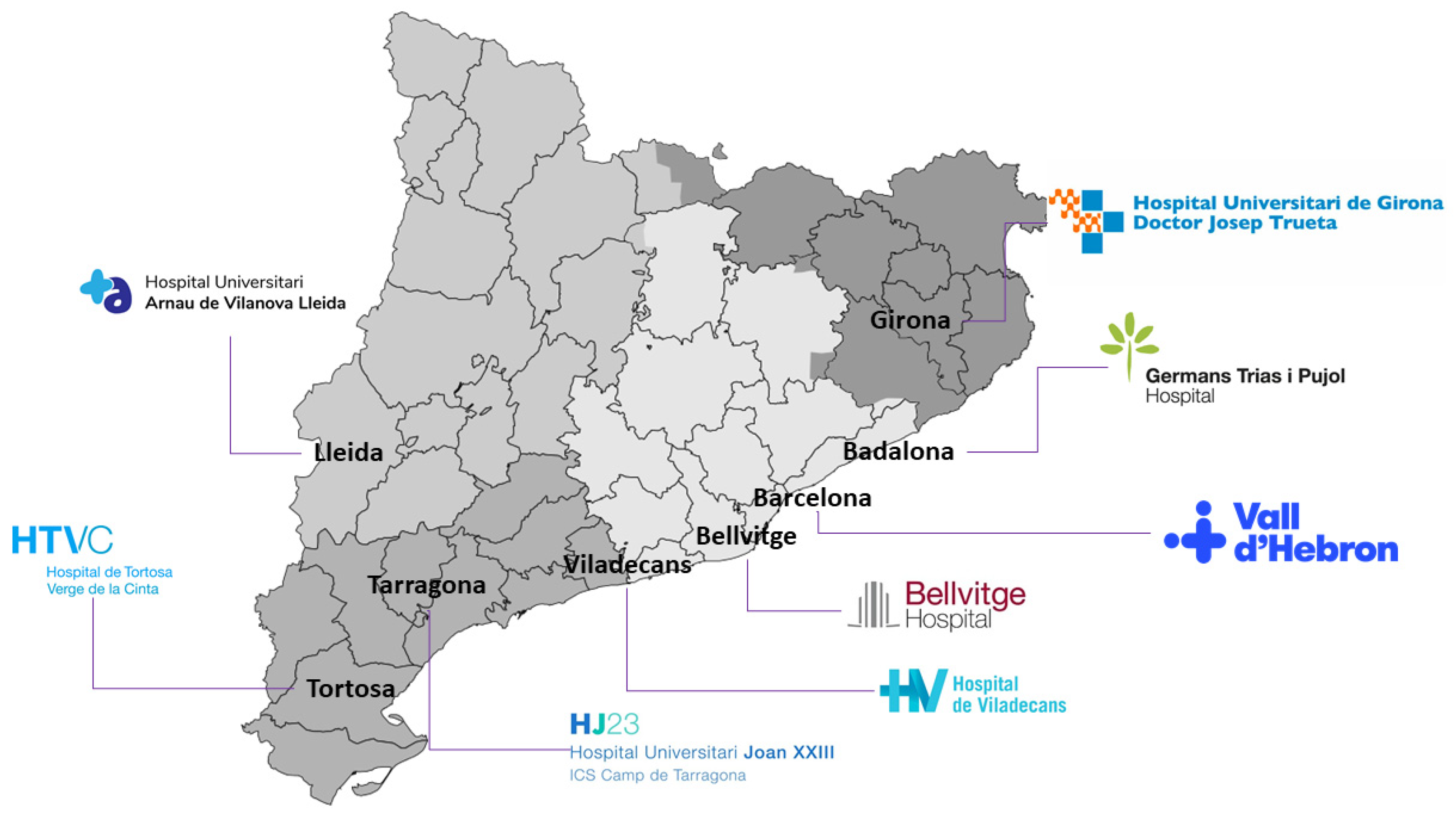
The Catalan Health Institute (Institut Català de la Salut or ICS) is the largest provider for the Catalan Health Service, the insurer of universal health coverage in Catalonia. It is the company with the most employees in Catalonia and the largest public company in Spain, with almost 39,000 professionals who provide services to almost six million people throughout the territory. [1] The ICS manages 283 primary care teams, three large high-tech tertiary hospitals (Vall d’Hebron, Bellvitge, and Germans Trias), four regional reference hospitals (Arnau de Vilanova in Lleida, Joan XXIII in Tarragona, Josep Trueta in Girona, and Verge de la Cinta in Tortosa), and a regional hospital (Viladecans) (Figure 1). The ICS accounts for seven percent of the Catalonian government budget, accounting for over 40 million primary visits and over 100,000 surgical interventions yearly.
|
Some laboratories are beginning to successfully deploy digital pathology solutions for routine diagnosis, which we believe will be a growing trend in the next few years. [2,3,4,5,6,7,8,9,10] In Catalonia, the DigiPatICS project plans to accomplish a complete digital pathology transformation of primary histopathological diagnosis for over 168 pathologists. Many groups have reported equivalency between digital pathology and conventional pathology. [11,12,13,14,15,16,17,18,19,20,21,22,23,24,25,26,27]
With the DigiPatICS project, we aim to increase patient safety and quality of care, improving diagnosis and the efficiency of processes in anatomical pathology departments of the ICS through digital pathology and artificial intelligence (AI) tools. [28] With digital pathology, we aim for a network of eight hospitals to work as one in terms of case sharing and teaching, putting all our patients on equal footing. This transversal digital transformation will have an impact on the care of patients treated by all medical and surgical specialists.
First, we created a network between ICS centers. This helped us increase the reproducibility and quality of diagnoses, as well as offered greater equity and safety to patients. In turn, this network approach facilitated remote diagnosis, case sharing, sub-specialization, and teaching for pathologists. In addition, we aimed for better working conditions, impacting the optimization of workflows, productivity, and, finally, turnaround times. We also intended to improve ergonomics and postural health, as well as to facilitate morphometric tools and the quantification of diagnostic and prognostic biomarkers to involve the optimization of time and a higher quality in diagnosis.
From a more technical point of view, the aim was to achieve a central digital repository of images on the network, thereby reducing the burden of slide file management and integrating medical imaging with SIMDCAT, a digital medical imaging system used in Catalonia. It was also intended as a subproject to establish bidirectional communications with other locations, such as operating rooms.
The project included the development of AI tools with machine learning (ML) and deep learning, taking advantage of the availability of whole slide images (WSIs) that were obtained after digitization. The objectives were to recognize tissue patterns, select tumor areas, and quantify them, among others. Hopefully, this use of AI tools will contribute to improving the quality of diagnosis and the efficiency of processes.
Materials and methods
DigiPatICS was created as a European Regional Development Fund (ERDF) project, with European funds for the optimization of anatomopathological diagnosis in a network of public ICS hospitals in Catalonia through digitalization and AI tools.
Subsequently, a market consultation was carried out, and, finally, it was tendered with the file code CSE/CC00/1101202869/20/AMUP [29].
Planning, scope, and tender process
A definition of needs was first carried out. We firmly believe meticulous planning is essential, taking into account all functional and technological requisites. Failing to detail such requirements can end in the failure of a digitization project, resulting in expensive scanners installed in pathology laboratories that are barely used. It is also important to highlight that going digital is not about acquiring pathology scanners; we focused our project on the purchase of a service with a shared risk with the bidder to achieve our objectives. Since this transformation was meant to be a one-way street with no possibility of going back to microscopes, all planning needed to include sufficient contingencies to avoid any kind of downtime for pathologists, as well as to ensure they benefit from the potential added value.
The purpose of the DigiPatICS project was to increase patient safety and quality of care, improving diagnosis and the efficiency of processes in pathological anatomy departments of the ICS using digital pathology and AI tools.
In defining the scope, several questions arose:
- Do we want to save the whole slide images (WSIs) forever? Who will store them?
- Is our laboratory information system (LIS) ready?
- Do we want to (or have to) address pre-analytics?
- Do we want to address dark-field microscopy (e.g., direct fluorescence, FISH)?
- Do we want to digitize the macroscopic images?
- Do we need to update the hospital network?
- Do we need to update our pathologists’ workstations?
- Do we want to share cases with the outside world?
- Is teaching important?
- Do we want AI algorithms?
- Do we want to do telepathology?
- What do we do with cytology?
- Do we have money for everything?
Those concerns and how they were resolved will be addressed shortly, but we can already answer some of these. We did want to store all the WSIs forever and to use that repository to train our own AI algorithms, which is clearly one of the great advantages of such a transformation. We also believed that this project must be an integral transformation, including routine histopathology, fluorescence, research, and macroscopic images. Tools for teaching and teleconsultation should be included, which meant having the option to share images outside our hospitals’ secured LAN. We realized that our LIS, preanalytics, network, and pathologist workstations all needed substantial upgrades to be able to undertake such a transformation. [9]
What about cytology? Digitizing cytology, even if feasible [30,31,32,33], has some particular concerns, including scanning times being much longer than in histology (due to the need for more resolution, larger scan area, zero tolerance of out-of-focus areas, and Z-stacking). That means needing to install more scanners to be able to take on the same activity, and it impacts storage needs. Dark-field scanning (FISH) has similar issues, but there was a significant difference in the volume of slides to scan. Cytology involves a large number of samples to digitize in our hospitals (over 400,000 each year), which is not affordable currently in this project due to budgetary constraints. (In other words, the activity addressed in our project included all bright-field, routine histopathology, histochemistry, immunohistochemistry, direct immunofluorescence, ISH, and FISH slides. Cytology was scanned on an as-needed basis.)
In Table 1, we summarize the total number of slides generated during 2019 at our eight hospitals, broken down by type. To that number of over one million slides, an expected growth in activity of 10% to 15% must be added each year. In addition, some resources were reserved for research and non-strictly routine samples and were not accounted for in these numbers.
| ||||||||||||||||||
Regarding the amount of personnel involved, DigiPatICS provided service to 107 pathologists, seven biologists, 40 residents, and 14 observers, adding up to a total of 168 professionals working with digital diagnosis.
In the tender process, all relevant aspects were taken into account for bidder evaluation, as shown in the following list:
- ▪ Automatic evaluation criteria (51 points)
- ○ Economic valuation (40 points)
- ▪ Evaluation of the financial offer (30 points)
- ▪ Evaluation of the maintenance offer (10 points)
- ○ Automatic technical evaluation (11 points)
- ▪ Quality management system. Certification of processes and algorithms (3 points)
- ▪ Process consulting (1 point)
- ▪ Image management platform adaptations (3 points)
- ▪ Storage for research slides (1 point)
- ▪ Short-term “hot” storage (3 points)
- ○ Economic valuation (40 points)
- ▪ Criteria subject to judgment value (49 points)
- ○ Scanners: deployment and image quality (17 points)
- ○ Diagnostic viewer (8 points)
- ○Image management platform (4 points)
- ○ Training module (2 points)
- ○ Built-in tools and algorithms (3 points)
- ○ Architecture and monitoring (1 point)
- ○ Definitive storage in SIMDCAT (4 points)
- ○ Integration of case information in a unified model (2 points)
- ○ Server infrastructure requirements and DPCs. Coherence, management model, virtualization (2 points)
- ○ Workstations (2 points)
- ○ Artificial intelligence (3 points)
- ○ Implementation and additional improvements (1 point)
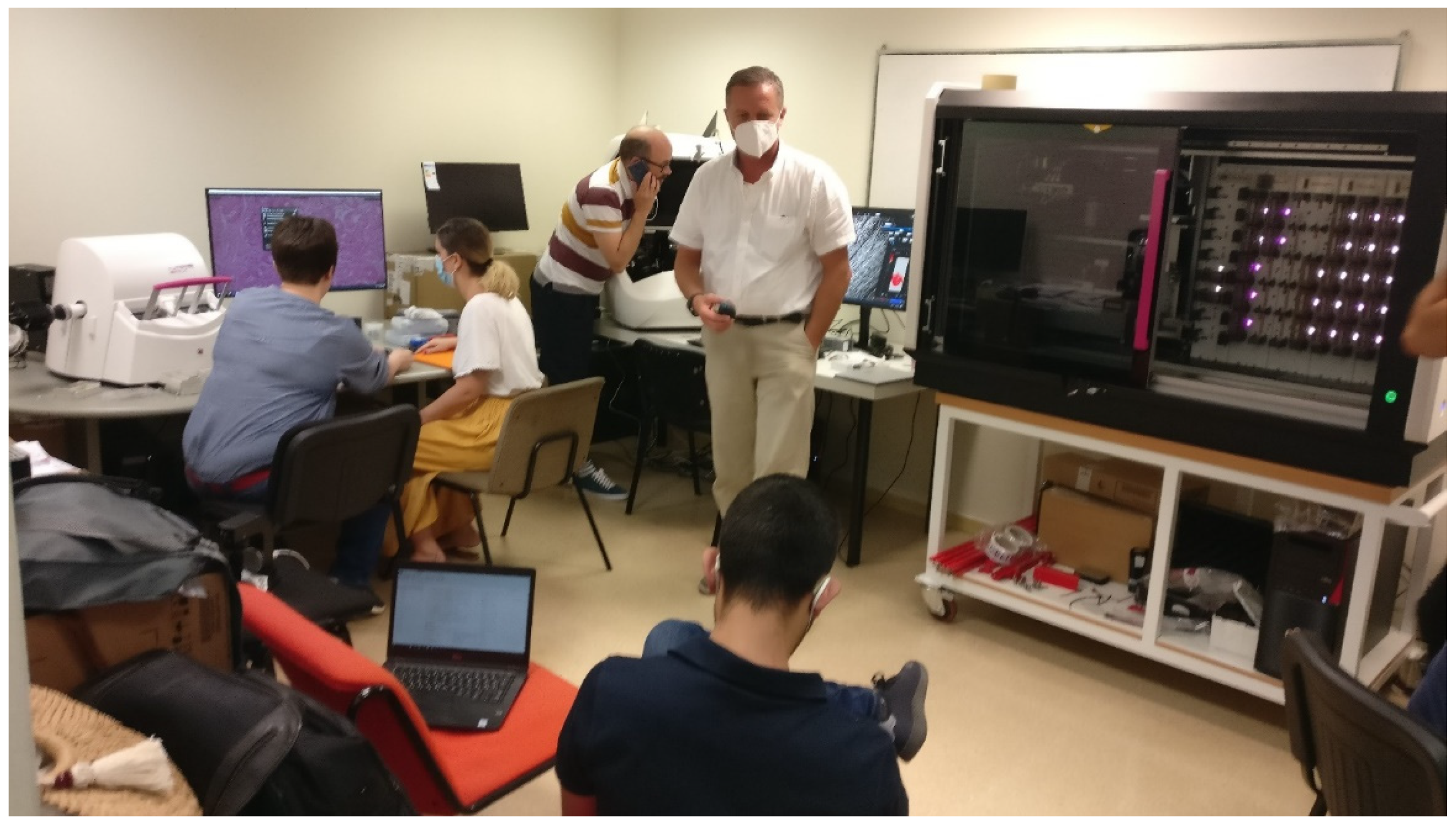
However, some aspects were considered very difficult to assess by evaluating their technical characteristics alone, and that is why three technical tests were defined for the scanner, viewer, and image management platform. For the scanner test, a large sample of glass slides from the eight hospitals was collected and fed to all the scanners offered by the three bidders for a week at 24 hours a day. In the fastest scanner, over 10,000 glass slides were digitized. Real scanning speeds, jamming, incidents, etc. were recorded to ensure the reliability of the equipment (Figure 2).
|
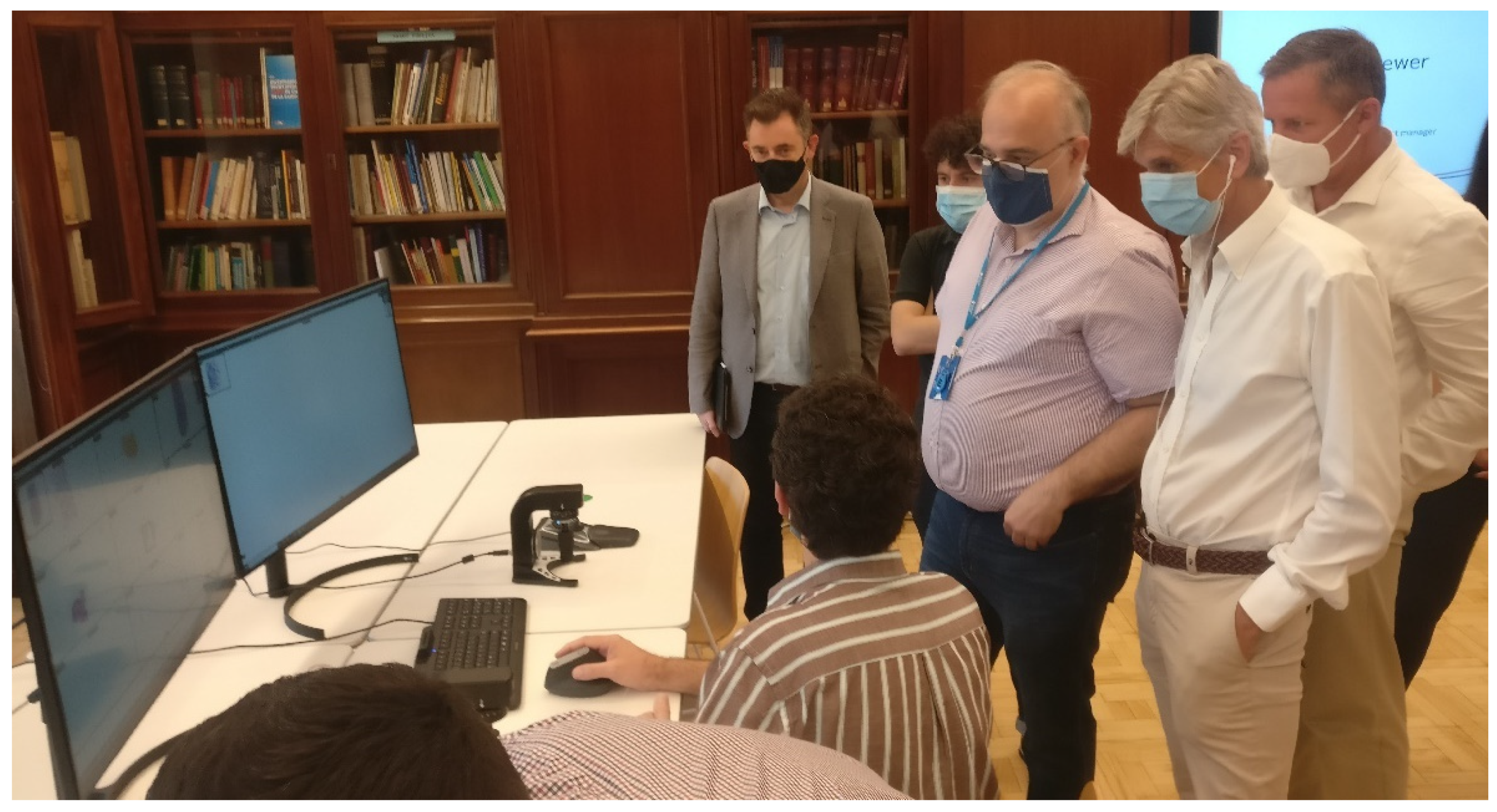
In the second and third tests, we brought together a group of pathologists from all the involved hospitals, along with IT experts, and they assessed the functionalities of the viewing software and the image management platform, as well as the image quality offered by the scanners using images scanned from ICS samples during the first technical test (Figure 3).
|
The contract file was definitively awarded by the decision of the Managing Director of the Catalan Health Institute (ICS) to the Palex Medical, S.A. 3DHISTECH Digital Pathology solution. This solution stood out for the proposal in the following main aspects:
- Scanners: Technical requirements, deployment requirements, and technical service requirements
- Image management platform: Diagnostic viewer, learning platform, and quantification modules
- Architecture: SIMDCAT interaction (DICOM), unified image management model (for all slide types), infrastructure management, and coherence model
Scanners and technology for obtaining whole slide images (WSIs)
Twenty-four scanners were installed and integrated into the workflow of the eight hospitals. Different scanner models were deployed according to the needs of each institution. In Table 2, we summarize the number of each scanner type and the capabilities.
| ||||||||||||||||||||||||||||||||||||||||||||||||||||||||
The PANNORAMIC 1000 Flash DX (3DHISTECH Ltd., Budapest, Hungary) (P1000) is a large (154 × 100 × 91 cm) and heavy (270 kg) scanner, but it offers the largest slide capacity on the market at 1,000 slides (using Leica slide racks, slide loading capacity could be further increased to 1,200). It is the fastest whole-slide scanner on the market, at up to 100 slides per hour and 2,000 slides per day (at 40× resolution, 0.25 μm/pixel, single layer). The P1000 uses Sakura slide racks, which seamlessly integrated with our laboratory workflow and allowed for priority slide handling and scanning in arbitrary order because it is flexible and automatic. It is also being used for double-width slides. Regarding image quality, it is able to scan at 0.25 μm/pixel, which is the 40× resolution equivalent (industry standard), and also at 0.12 μm/pixel, which is roughly the 80× resolution equivalent. Multilayer (Z-stack) and extended focus scanning are available, as well as automatic water immersion. [34,35] Furthermore, thanks to its AI-based software control, it is able to automatically rescan suboptimal slides, adding multilayer scanning if required. The P1000 are used for all bright-field and double-width slide related imaging.
The PANNORAMIC 300 Flash DX (3DHISTECH Ltd., Budapest, Hungary) (P300) is a fast bright-field and fluorescence scanner capable of high throughputs as a standalone machine in smaller institutions or serving as backup for P1000s in larger hospitals. It has the capacity for 300 slides, and its use is mainly for fluorescence, scanning FISH, and direct immunofluorescence. [36]
Both the PANNORAMIC SCAN II and the PANNORAMIC MIDI (3DHISTECH Ltd., Budapest, Hungary), with slide capacities of 150 and 12 slides, respectively, are mainly focused on fluorescence imaging but are still able to scan bright-field images, even though they are slower than their high-throughput counterparts. [37]
Macroscopic imaging
Regarding macroscopic imaging, 13 MacroPATH QX systems (Milestone Medical, Sorisole, Italy) were installed to obtain and incorporate gross imaging into the workflow. All the images were stored on the DigiPatICS servers and were fully integrated and available at the pathologist workstations for making diagnoses.
WSI viewing: Hardware
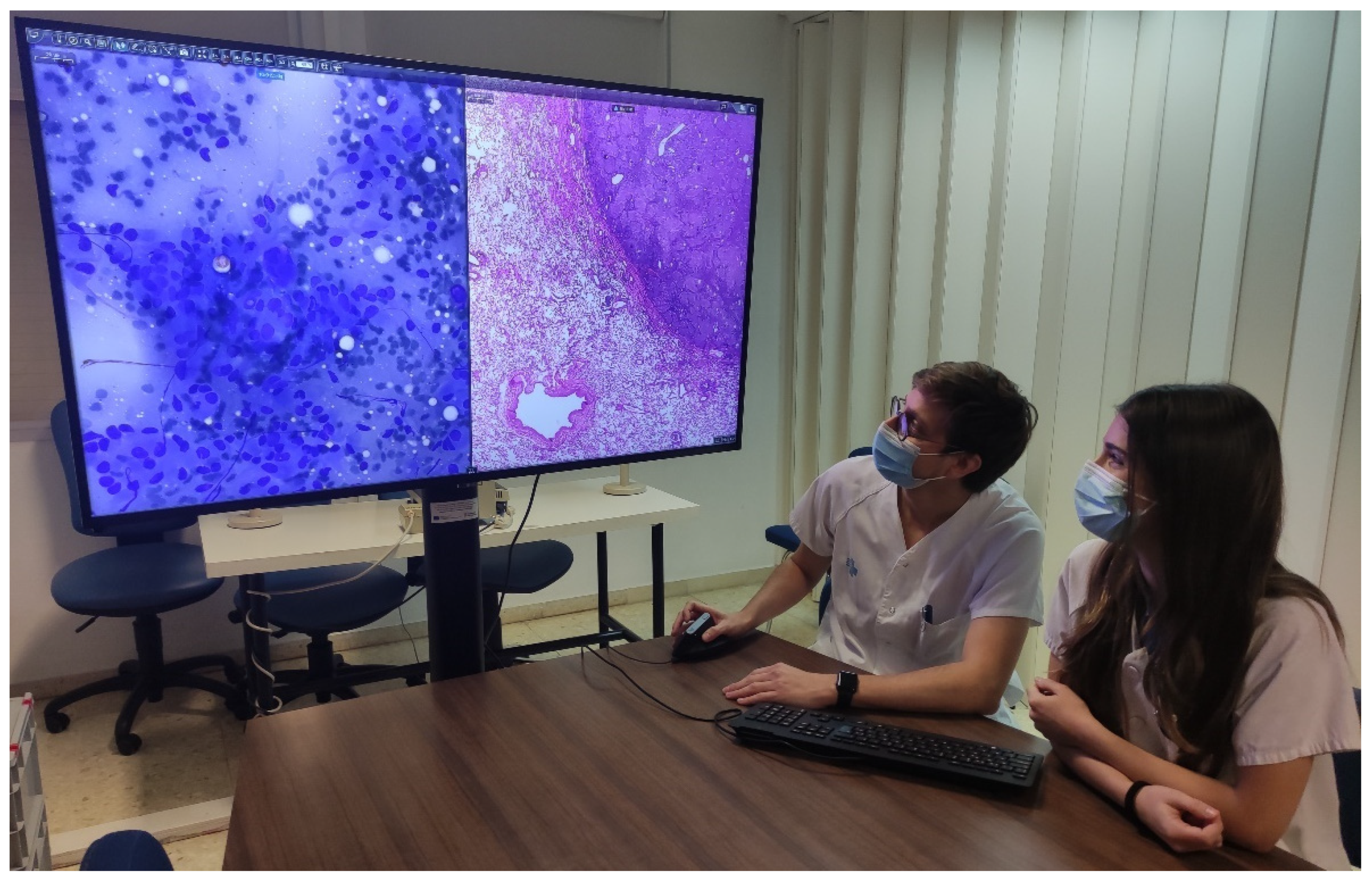
To be able to view WSIs, 183 new workstations were installed (Table 3) for pathologists, residents, biologists, observers, and meeting rooms. Each pathologist workstation consisted of two 32-inch 4K UHD (3840 × 2160 pixel) diagnostic medical-grade FDA-approved monitors (LG 32HL512D) (Figure 4) that could be used indistinctly in flexible ways. Normal intended use is for a pathologist to have an LIS with all laboratory data, clinical data, and reporting available on one monitor, while on the other, a microscopic image is displayed. However, both monitors could be used for microscopic images, or both could be used for reporting, clinical data, bibliography, or other tasks. Biologist workstations were the same as those of the pathologists. Residents and observers shared the same workstations; however, they only consisted of one LG 32HL512D medical-grade monitor due to space constraints (the dual-monitor setup required over 150 cm of desk surface).
| ||||||||||||
|
Thirteen additional workstations with six 55-inch 4K UHD monitors and seven 4K UHD DICOM ProBeam LG projectors were installed in small and medium meeting rooms for teaching and clinical sessions as a replacement for multi-head microscopes (Figure 5).
|
Each workstation was comprised of an Intel Core i5-9600K processor (Intel Corporation, Santa Clara, CA, USA), 16 Gb RAM, 512 Gb SSD, and an RTX 2060 graphics card (Nvidia Corporation, Santa Clara, CA, USA). All workstations were the same for easier maintenance, compatibility, and interchangeability of workplaces. Each workstation also contained a Logitech BRIO 4K UHD webcam (Logitech International, Lausanne, Switzerland) and a Jabra Evolve 40 headset with a microphone (GN Group, Ballerup, Denmark). Both the webcam and microphone aimed to facilitate networking among pathologists from the same hospital, from different hospitals within the ICS, or even with professionals outside our network.
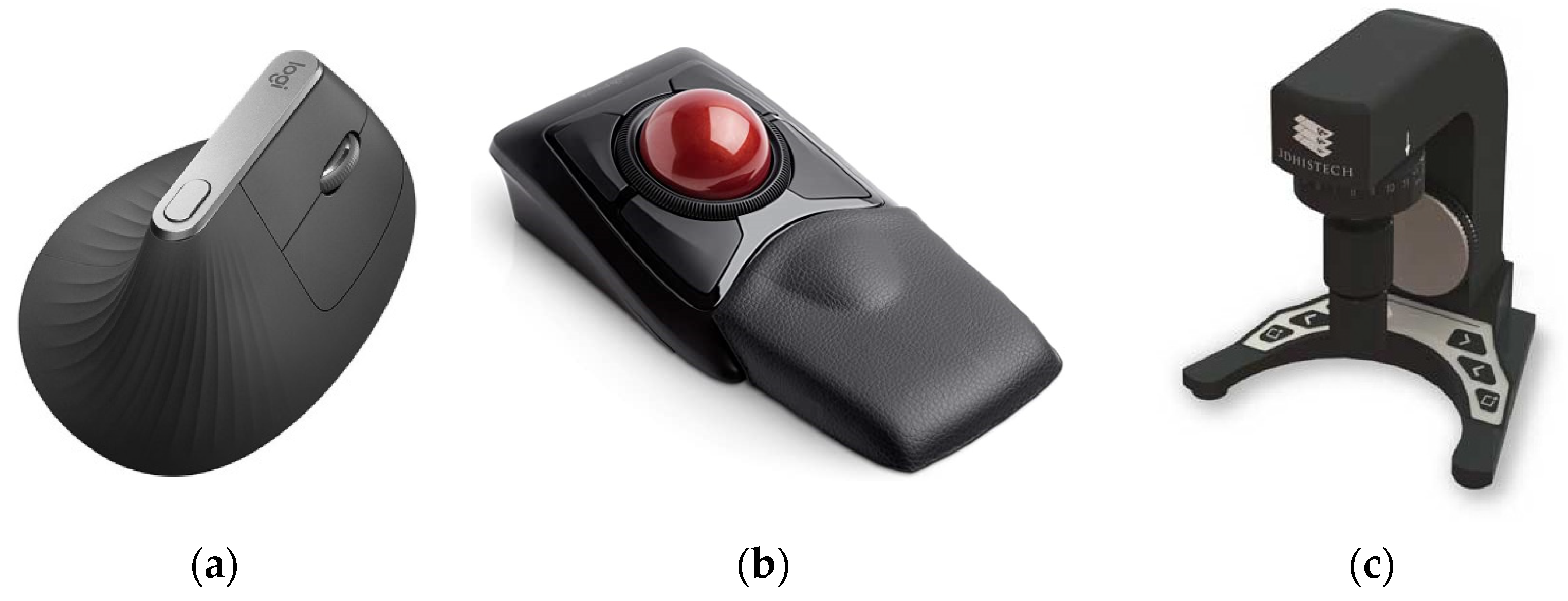
Furthermore, each workstation contained a Logitech MX Vertical Ergonomic Wireless Mouse (Logitech International, Lausanne, Switzerland), since vertical mice seem to put less strain on the wrist and demonstrate better ratings than conventional mice. [38] Each pathologist could also choose between two other ergonomic devices: a Kensington Expert Mouse Wireless Trackball (Kensington Computer Products Group, Redwood Shores, CA, USA) and a SlideDriver (3DHISTECH Ltd., Budapest, Hungary) (Figure 6). All devices were supported by our viewing software. The SlideDriver offers microscope-like navigation on digital slides for those who prefer a traditional method. Most of our pathologists and residents selected the SlideDriver as their input device (80% approximately).
|
WSI viewing: Software
The diagnostic viewer used for all digital images was ClinicalViewer (3DHISTECH Ltd., Budapest, Hungary). It uses streaming technology to avoid downloading WSIs for diagnosis. It is capable of opening bright-field, fluorescence, double-slide, Z-stack, and macroscopic photography, etc. It also includes many positively valued features, such as the possibility of viewing and navigating up to nine automatically synchronized images at once. It also has IVD support and quantification algorithms, as well as some more standard tools, such as free rotation, free zoom, annotation, measuring, and object counting tools (Figure 7).
|
The software also had a telepathology option available and a module to create training courses. Possessing all the WSI viewing needs, including fluorescence, integrated into one software system (one viewer) facilitated pathologist ergonomics and also enabled simplifying the technological model with fewer integrations required.
The software also had a telepathology option available and a module to create training courses. Possessing all the WSI viewing needs, including fluorescence, integrated into one software system (one viewer) facilitated pathologist ergonomics and also enabled simplifying the technological model with fewer integrations required.
ETPAT: Our laboratory information system (LIS)
The evolution of information systems in the last 10 years has been meteoric. It has changed the paradigms for accessing and possessing the necessary information at each point of contact a patient has with a health system.
References
Notes
This presentation is faithful to the original, with only a few minor changes to presentation, though grammar and word usage was substantially updated for improved readability. In some cases important information was missing from the references, and that information was added. At the Catalan Health Institute (ICS), and within the ARGOS project, we have spent 15 years directing information towards users and clinical care, and we have progressively moved from the initial free texts to structured information. The ARGOS project started in 2006. It is a project to integrate in a transversal and transparent way all the information systems involved in clinical assistance to the citizens of the Catalan Health Institute and its eight hospitals, including the hospital information system, nursing, pharmacy, clinical analysis laboratory, pathology laboratory, and critical care units. Currently, ARGOS is the priority information system in Catalonia and is present in 23 hospitals of the Catalan public health network.
The SALUT4D project began in 2020 and is the evolution of clinical workstations within the ARGOS project. Its objective is to provide the necessary information at each moment of care to different professionals. It is based on four dimensions seeking to present the necessary data at each point of care:
- Where am I? Scope of work, including emergencies, hospitalization, ambulatory consultation, operating room, etc.
- Who am I? Role, including nurse, surgeon, internist, psychologist, etc.
- Whom do I attend? A patient with hypertension, diabetes, bronchitis, etc.
- How do I attend to it? With a computer, tablet, smartphone, etc.
The system presents the information that a professional needs clearly and orderly. The system is based on a clinical dictionary with more than 40,000 variables stored in a MongoDB-type database called the Global Variables Repository (RGV). This repository contains data from all sources, including laboratory, pathological anatomy, radiology, vital sign monitors, pharmacy, etc.
To construct this platform, created and designed for and by the ICS, HTML5 technology, SAP ISH (Information System Hospital), and SAP ISHMed (SAP SE, Walldorf, Germany) were used. The system connects directly with the Shared Clinical History of Catalonia (HC3) and provides access to information on all public hospitals and all primary care facilities in a fully integrated manner.
The workflow methodology was based on requirements submitted by over three hundred health professionals who continuously provided their contributions. Through AGILE methodologies, the new clinical workstation was progressively built. The new station is already in operation at the eight ICS hospitals and will gradually come into operation in 23 sections of the ARGOS system throughout 2022.
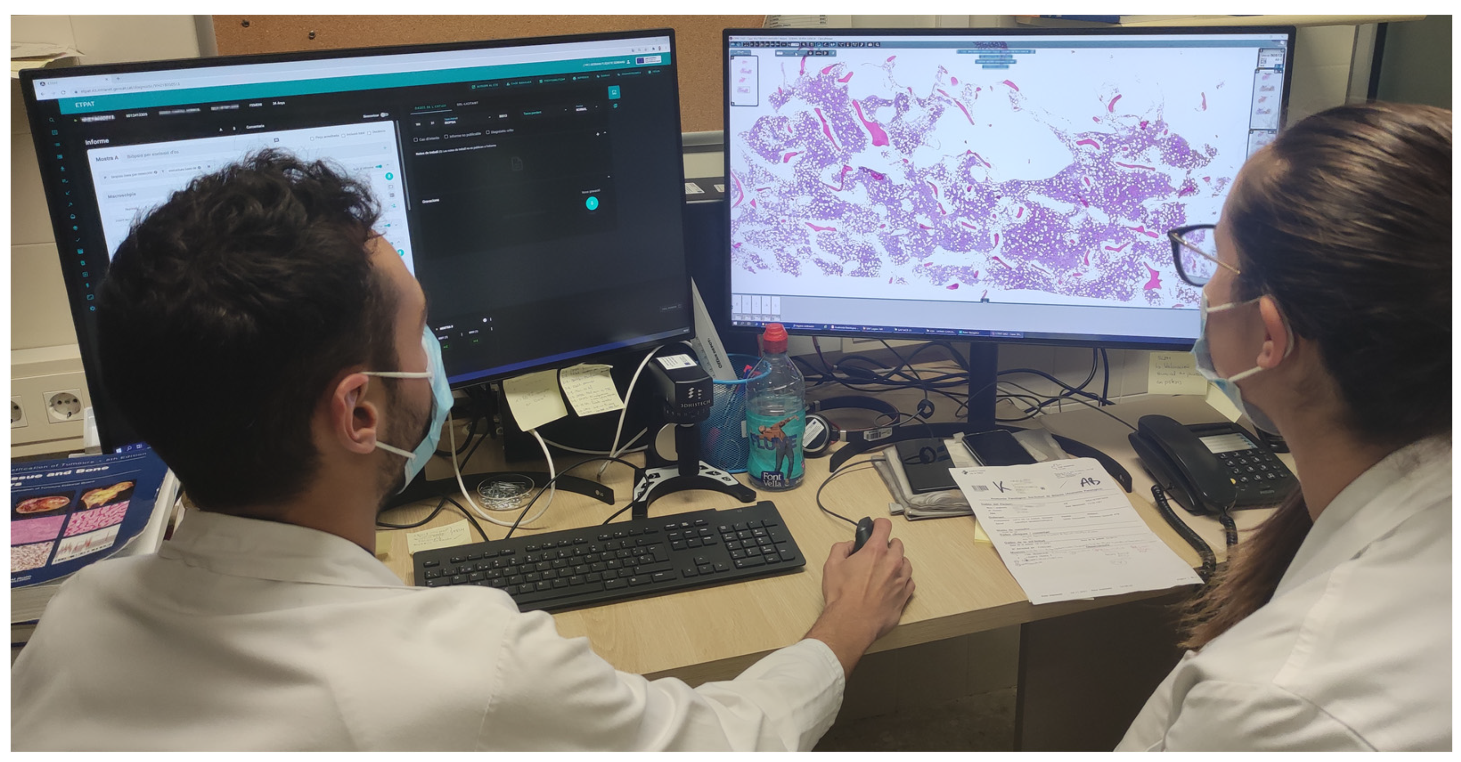
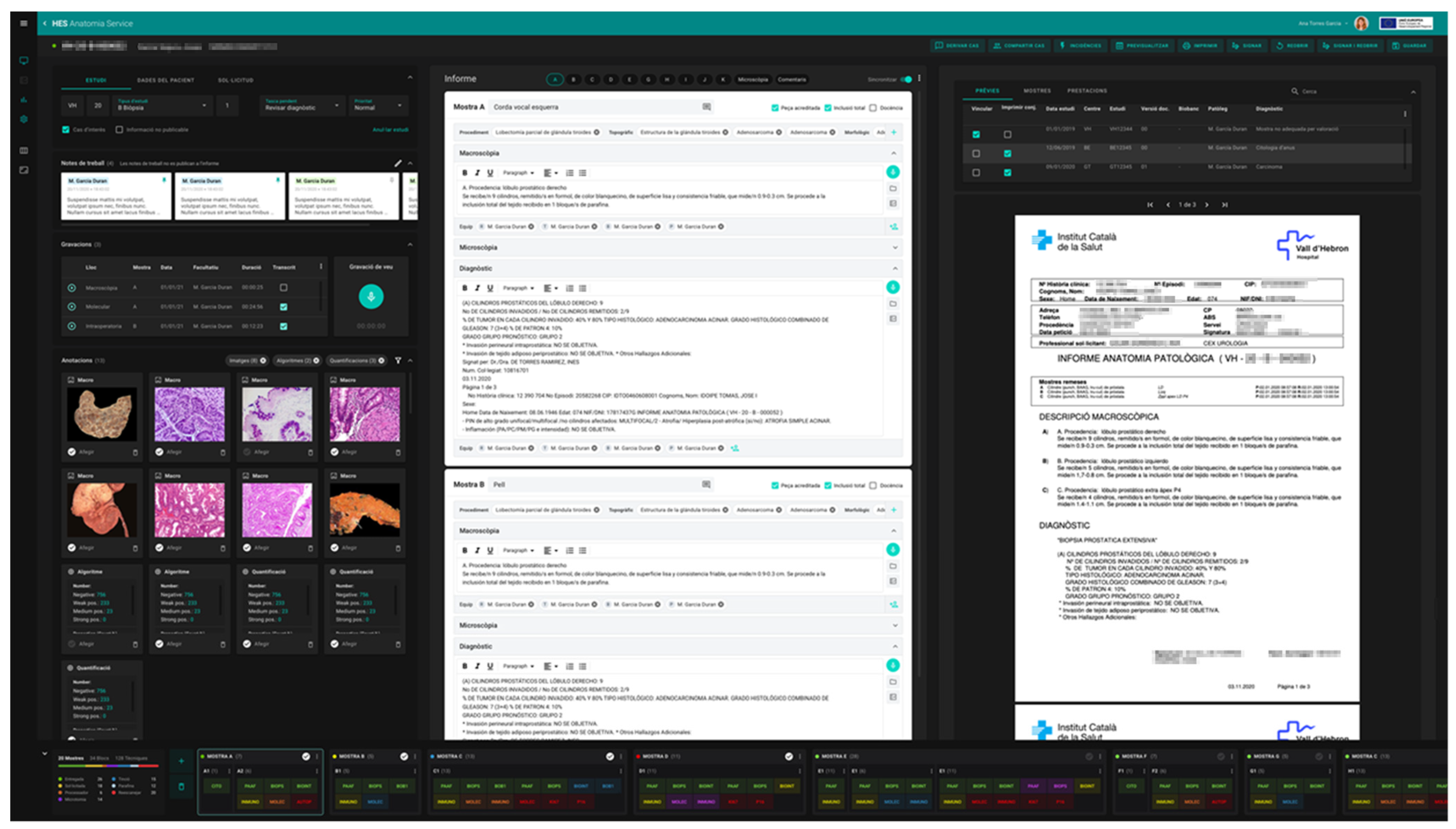
In order to incorporate the peculiarities of the new, fully digital workflow, a new LIS for our pathology laboratories named ETPAT was developed. It was deemed necessary to develop and improve pathologist workstations to provide them with tools that allow the integration of information from all sources and electronic records available. ETPAT has a fully integrative approach to all processes occurring in our pathology labs, from electronic requests from clinicians to pathology reports, with full traceability of all steps involved at the laboratory. Moreover, it enables the optimization of paper management as much as possible. This software was based on HTML5 and was prepared for 32-inch 4K UHD monitors, taking advantage of the hardware on hand (Figure 8).
|
